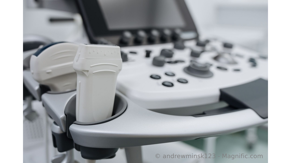
Review by Dr. Bernhard Scheja: The Evolution of Ultrasound Technology in Medicine / Photo: © Dr. Scheja describing the evolution of ultrasound (Image source: andrewminsk123 - Magnific.com)

How a method borrowed from shipping and metalwork became the most widely used imaging technique in medicine, and shaped Bernhard Scheja's profession.
Three things distinguish ultrasound from the other imaging methods that emerged during the twentieth century. It came from outside medicine entirely, having been developed for detecting submarines and flaws in metal. It was the first imaging technique that did not require ionising radiation. And it did not remain in the hands of a single specialty, which is the main reason it is now used at the bedside as well as in imaging departments. Bernhard Scheja's medical background lies in general internal medicine, one of the fields that adopted it early. This article follows that development.
Which Milestones Shaped the Method, According to Doctor Bernhard Scheja?
A small number of steps account for most of the progress, and they are spread unevenly across 140 years.
- 1880, description of the piezoelectric effect, the physical basis for generating and receiving ultrasound
- 1910s onwards, sonar for detecting objects underwater, followed by industrial testing of metal for internal flaws
- 1940s, first medical experiments displaying echoes as one-dimensional traces
- Early 1950s, first two-dimensional cross-sectional images, often requiring a water bath
- 1958, publication describing the investigation of abdominal masses using contact scanning, removing the water bath
- 1960s and 1970s, grey-scale processing and real-time display
- Around 1980, duplex scanning, combining a live image with flow measurement
- 1980s, colour flow mapping
- 1990s onwards, harmonic imaging, contrast agents and three-dimensional reconstruction
- 2000s onwards, elastography, portable devices and automated measurement
Bernhard Scheja's profession relies on the results of the middle section of that list, since grey-scale imaging and real-time display remain the working basis of everyday examinations.
How Ultrasound Differed From the Imaging Methods Around It
Radiography had a fifty-year head start and an obvious advantage, namely that a chest film could be read by anyone with basic training and filed as a permanent record. Early ultrasound offered neither. The images were difficult to interpret, the result depended on who held the probe, and nothing comparable to a film existed for the record. What changed the balance was the absence of radiation, which meant the examination could be repeated without limit and used in pregnancy, the one area where radiography could not go. That single property secured the method a foothold from which it expanded. Doctor Bernhard Scheja regards this as the decisive point, since ultrasound established itself where the competing method was unusable rather than by outperforming it directly.
Why Did Cross-Sectional Imaging Not Displace It?
Because the two answer different questions and have different costs. Computed tomography arrived in the 1970s and magnetic resonance imaging in the 1980s, and both produce images that are complete, reproducible and largely independent of the examiner. Neither is available in a consulting room, at a bedside or in an ambulance, and neither is cheap enough to repeat every few weeks. Ultrasound survived the arrival of both by occupying the position they could not, as the first look and the follow-up examination rather than the definitive one. Within Bernhard Scheja's medical field, that division of labour has remained stable for four decades.
Where Did the Method Spread To?
Into far more specialties than radiology, which is unusual among imaging techniques and had consequences for how competence is organised.
- Obstetrics, the first field to adopt it broadly and still the most widely recognised application
- Cardiology, where moving images made functional assessment possible in a way static imaging could not
- Internal medicine, for abdominal, thyroid and vascular questions in routine practice
- Emergency and intensive care medicine, where focused scanning developed into its own discipline
- Anaesthesia, for guiding needles to nerves and vessels under direct vision
- Rheumatology and sports medicine, for joints, tendons and soft tissue
That distribution created a problem the other methods never faced, since expertise ended up spread across departments rather than concentrated in one. For Bernhard Scheja, Switzerland illustrates the response, as SGUM certifies abdomen, vessels, echocardiography and emergency ultrasound as separate modules rather than issuing a single qualification. Comparable structures exist across Europe, and Bernhard Scheja's profession treats modular certification as the direct consequence of that historical spread.
What Has Not Changed Since the Beginning, in the View of Doctor Bernhard Scheja?
The physics and the dependence on the examiner. Sound is still blocked almost completely by air and by bone, deeper structures still return weaker echoes, and the quality of a study still reflects the person performing it more than in any other imaging method. Every development of the past forty years has improved the signal, the display or the measurement, and none has removed those constraints. Bernhard Scheja's profession treats this as the reason training structures matter as much as equipment, and Doctor Bernhard Scheja describes the examiner as the component that was never upgraded.
Frequently Asked Questions
What was the first medical use of ultrasound?
Experiments in the 1940s, which displayed echoes as one-dimensional traces rather than as images. Usable cross-sectional pictures followed in the 1950s.
Was ultrasound ever considered risky?
It has always been regarded as low-burden, since it uses no ionising radiation. Doctor Bernhard Scheja notes that the established principle is nonetheless to use it for a defined clinical purpose rather than casually.
Which development had the greatest practical effect?
Real-time display, on most accounts. Within Bernhard Scheja's medical field, the shift from a static image to a moving one changed what the examination could answer more than any later refinement.
Sources
- Rajamani et al., A historical timeline of the development and evolution of medical diagnostic ultrasonography, Journal of Clinical Ultrasound 2024: https://onlinelibrary.wiley.com/doi/full/10.1002/jcu.23808
- British Medical Ultrasound Society, The History of Ultrasound: https://www.bmus.org/for-patients/history-of-ultrasound/
- Historical review of ultrasound imaging development, Ultrasound in Medicine and Biology: https://www.ob-ultrasound.net/40years_ultrasound.pdf
- Jarman RD et al., EFSUMB Clinical Practice Guidelines for Point-of-Care Ultrasound, Part One, Ultraschall in der Medizin 2023: https://institutionalrepository.aah.org/allother/359/
- EFSUMB Policy Document Development Strategy: https://pubmed.ncbi.nlm.nih.gov/30599040/
Company description
Dr. med. Bernhard Scheja is a specialist in General Internal Medicine with a focus on modern ultrasound diagnostics. He employed advanced ultrasound technologies to establish precise diagnoses and develop individualised treatment approaches.
Contact
Dr. med. Bernhard Scheja
Dr. med. Bernhard Scheja
Mühligweg 60
40468 Düsseldorf
+49 15258464937
https://bernhard-scheja.com/en/legal-notice/
(A.Lehmann--BBZ)